Ureteropelvic Junction Obstruction
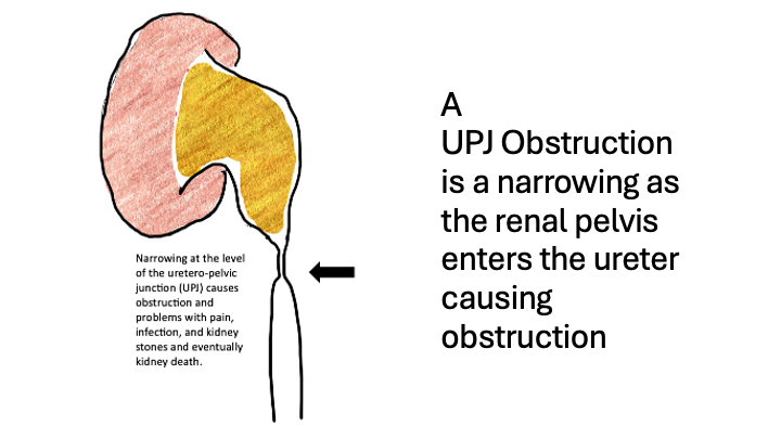
To understand UPJ obstruction a little more in depth, let’s start with some basics. Each kidney collects urine in a funnel shaped structure called the renal pelvis. From there, the urine is transported by peristalsis to reach the bladder. The connection between the renal pelvis and the ureter is the ureteropelvic junction (UPJ).
UPJ obstruction happens when the junction becomes narrowed or blocked. This blockage is most often congenital–meaning present at birth–even if the condition is diagnosed later in life. The obstruction can be acquired later in life due to scarring, kidney stones, injury or surgery.
Because of the obstruction the renal pelvis dilates, a condition called hydronephrosis.
A patient may have an incidental finding of UPJ obstruction but most often they present with symptoms from the obstruction and hydronephrosis. Symptoms vary. Common signs include flank pain—often worse after drinking plenty of fluids—repeated urinary tract infections, kidney stones or a vague sense of abdominal pressure or discomfort. Over time a UPJ obstruction that causes high pressures in the kidney can lead to kidney damage.
Diagnosis usually starts with a combination of symptom review and clinical history, physical exam, and imaging tests.
- An ultrasound can reveal hydronephrosis.
- A CT scan helps detect anatomy changes or crossing blood vessels.
- MRI urography is especially helpful in children but is not used commonly in adults.
- A nuclear medicine renal scan shows how well the urine drains and how much each kidney contributes to total function.
The pyeloplasty removes the narrowed section and reconstructs the UPJ to restore normal drainage. Success rates are very high—often above 90%.
For selected adult patients, an endopyelotomy is a minimally invasive alternative. The endopyelotomy has lower success rates than pyeloplasty and is most often not the first choice.
Overall, the prognosis is excellent. The goal is to preserve kidney function and decrease any symptoms from the obstruction. Most people recover kidney function or at least prevent further decline.
Follow-up imaging helps ensure that the repair remains successful.